Skip to content
Human Physiology · Biology

Human Heart & Double Circulation 3D Simulation

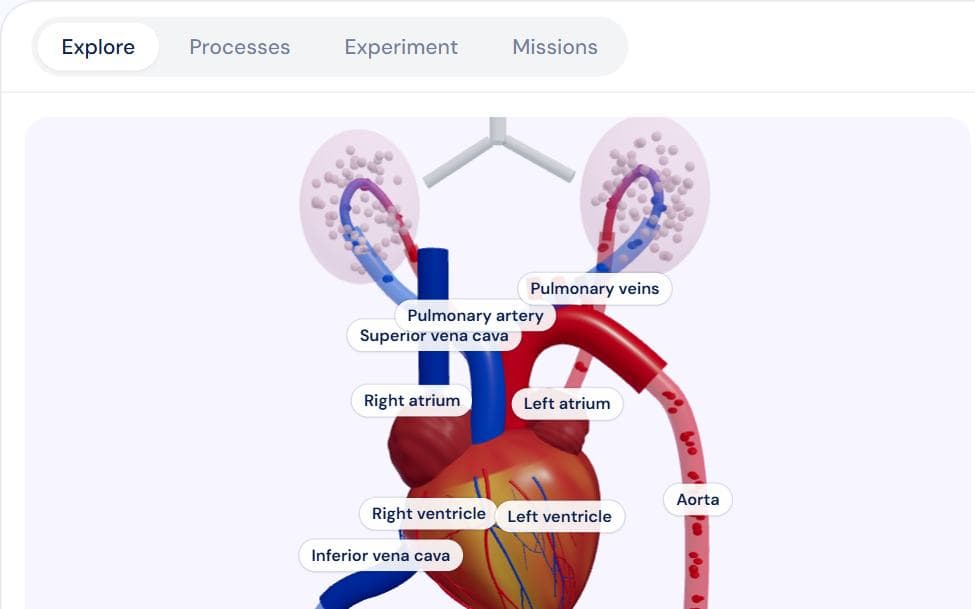
A beating 3D heart wired to the lungs and the body, with blue and red blood cells flowing round both circuits. Click any chamber, valve or vessel; play double circulation, the cardiac cycle (with a live ECG and lub-dub) and the conduction system step by step; change the heart rate or add a defect; then trace a red cell and answer NEET questions.

Interactive virtual lab · no download · works on phones and laptops

Chapter
Body Fluids & Circulation
For
Class 10 · Class 11 · NEET
Time
~25 min

Human Heart & Double Circulation 3D Lab is a Pro simulation

Pro opens all 152 virtual labs — 693 simulations with guides and quizzes.

How to use it

  1. 1Explore: drag to turn the heart, click a part; use the circuit buttons to show only the pulmonary or systemic loop.
  2. 2Processes: play double circulation, the cardiac cycle or the conduction system step by step.
  3. 3Experiment: pick an activity or set rate and stroke volume; add a defect, predicting first.
  4. 4Missions: find parts, trace a red cell round the body, and NEET questions.

What to notice

  • One lap passes through the heart twice: right side → lungs → left side → body. That is double circulation.
  • The pulmonary artery carries oxygen-poor blood and the pulmonary veins oxygen-rich blood: arteries leave the heart, veins return to it.
  • 'Lub' is the AV valves shutting as the ventricles contract; 'dub' is the semilunar valves shutting as they relax.
  • P wave → atria contract, QRS → ventricles contract, end of T → end of systole.
  • Cardiac output = heart rate × stroke volume ≈ 72 × 70 mL ≈ 5 L/min at rest.

For teachers

A ready-made teaching kit comes with this lab

  • Lesson plan

    28 min, 4 steps

  • Ask the class

    5 questions with answers

  • Quick quiz

    6 questions, printable

  • Key formulas

    4 formulas

About the Human Heart & Double Circulation 3D Lab simulation

This 3D lab shows the human heart the way a textbook diagram of double circulation does, but beating. Four chambers contract in the right order, the tricuspid, bicuspid and semilunar valves open and shut on cue, and blood cells flow from the venae cavae through the right side of the heart to the lungs, back through the left side and out through the aorta to the body organs. Cells are blue while they carry oxygen-poor blood and turn red in the lung capillaries, then blue again in the body's capillaries, so the two circuits are easy to follow.

The lab has four modes. Explore lets you click any chamber, valve, vessel, the lungs, the body organs or the conduction system to read its structure, function and the points NEET asks about, with a live ECG strip that marks the phase, the valve states and each 'lub' and 'dub'. Processes plays three sequences step by step: double circulation (nine steps, following a single red cell), the cardiac cycle (each phase in slow motion) and how each beat starts (SA node, AV node, bundle of His, Purkinje fibres). Experiment changes heart rate and stroke volume to show cardiac output, and adds a hole in the ventricular septum or a leaky bicuspid valve. Missions ask you to find parts, trace a red cell round the body by clicking each stop, and answer NEET questions.

The numbers follow NCERT's Body Fluids and Circulation: about 72 beats a minute, a cycle of about 0.8 seconds split into 0.1 s atrial systole, 0.3 s ventricular systole and 0.4 s joint diastole, a stroke volume of about 70 mL and a cardiac output of about 5 litres a minute. The defects are an illustrative model that shows the direction of the change, not clinical values.

Also covers: human heart 3d, double circulation, pulmonary circulation, systemic circulation, heart chambers, right atrium, left ventricle, tricuspid valve, bicuspid valve, mitral valve, semilunar valve, pulmonary artery, pulmonary vein, aorta, vena cava, cardiac cycle, lub dub, heart sounds, ecg, p wave qrs t wave, sa node pacemaker, av node, bundle of his, purkinje fibres, cardiac output, stroke volume, neet body fluids and circulation, class 10 life processes.

Frequently asked questions

What is double circulation?

In one complete lap blood passes through the heart twice: the right side pumps oxygen-poor blood to the lungs (pulmonary circulation), and the left side pumps the oxygen-rich blood that returns from the lungs to the rest of the body (systemic circulation). A complete septum keeps the two kinds of blood apart, so the heart works as a double pump.

Why does the pulmonary artery carry deoxygenated blood?

Arteries are named for the direction of flow, not for their oxygen: they carry blood away from the heart. The pulmonary artery takes blood from the right ventricle to the lungs, before it has picked up oxygen. In the same way the pulmonary veins carry oxygen-rich blood, because they bring it back to the heart from the lungs.

What causes the 'lub' and 'dub' heart sounds?

'Lub', the first sound, is the tricuspid and bicuspid valves shutting at the start of ventricular systole. 'Dub', the second sound, is the semilunar valves shutting at the start of ventricular diastole. Between them, in ejection, the semilunar valves are open and the AV valves are shut.

What do the P, QRS and T waves of an ECG show?

The P wave is the depolarisation of the atria, which makes them contract. The QRS complex is the depolarisation of the ventricles and starts ventricular contraction. The T wave is the ventricles returning to normal (repolarisation); its end marks the end of systole. Counting QRS complexes in a minute gives the heart rate.

Why is the wall of the left ventricle thicker than the right?

The left ventricle pumps blood round the whole body against a high resistance, while the right ventricle only pumps it through the nearby lungs. The left side has to develop a much higher pressure, so its muscle is thicker.